New Arrivals
Deals & Promotions
Patterson Private Brand
Patterson Office Supplies
Browse by Manufacturer
Controlled Substances
Custom Products & Services
3M
Dentsply Sirona
Kavo
My Catalog
All Merchandise Categories
Adhesive Agents, Materials & Accessories
Adhesive Agents, Materials & Accessories
Air/Water Syringes & Evacuation System Parts
Air/Water Syringes & Evacuation System Parts
Air/Water Syringe & Evacuator Assembly Replacement Parts
Air/Water Syringe & Evacuator Assembly Replacement Parts
CAD/CAM Blocks & Accessories
CAD/CAM Blocks & Accessories
CAD/CAM TiBase Kits, Drill Keys & Implant Scanning
CAD/CAM TiBase Kits & Abutment Screws
CAD/CAM Scan Posts
CAD/CAM Scanbodies
CAD/CAM Drill Key Sets
CAD/CAM Guide Block Kits
CAD/CAM Training Models & Model Fabrication Accessories
CAD/CAM Model Holders, Baseplates & Adapters
CAD/CAM Training & Calibration Models
CAD/CAM Model Fabrication Kits
Cotton & Paper Supplies
Cotton & Paper Supplies
Cotton Rolls, Pellets & Sponges
Absorbent Sponges
Cotton Pellet Dispensers
Cotton Pellets & Alternatives
Cotton Roll Dispensers & Holders
Cotton Rolls & Alternatives
Sponge Dispensers
Tongue Depressors
Sundry Jars & Holders
Crown & Bridge
Dispensing Syringes, Guns & Mixing Supplies
Dispensing Syringes, Guns & Mixing Supplies
Automatic Mixers & Vacuum Mixing Bowls
Automatic Mixing Unit Replacement Parts
Automatic Mixing Units
Mixer Mounts & Stands
Vacuum Mixing Bowls & Paddles
Dispensing Guns, Syringes & Applicators
Cotton-Tipped Applicators & Alternatives
Applicator Brushes, Handles & Tips
Dispensing & Irrigating Syringes
Dispensing Guns
Gun & Syringe Replacement Parts
Gun & Syringe Stands
Gun & Syringe Tips, Needles & Caps
Mixing & Intraoral Cartridge Tips
Pipettes & Droppers
Endodontics
Endodontics
Apex Locators & Obturation Systems
Obturators
Apex Locators, Pulp Testers & Accessories
Obturation Systems & Accessories
Endodontic Points and Medicaments
Gutta Percha Points
Absorbent Paper Points
Silver Points
Endodontic Medicaments
Endodontic Files, Reamers & Accessories
Endodontic Shaper
Endodontic Hand Files
Endodontic Rotary Files
Endodontic Reamers
Paste Fillers
Broaches
Endodontic Drills
Endodontic Pluggers & Spreaders
Endodontic Test Handles
Endodontic Gauges & Rulers
Endodontic Stops
Endodontic Organizers & Accessories
Endodontic Miscellaneous
Finishing & Polishing
Front Office Products
Front Office Products
Appointment & Scheduling Products
Appointment Book Accessories
Dated Wirebound Appointment Books
Undated Wirebound Appointment Books
Standard Appointment Cards
Sticker Appointment Cards
Undated Looseleaf Sheets & Binders Appointment Books
Scheduling Systems
Billing Products & Daily Log
Miscellaneous Billing Products
Laser Checks
Manual Checks
Daily Log
ADA & HCFA/CMS Insurance Forms
One Write Accessories
One Write Products
Receipts and Deposit Slips
Laser Statements
Manual Statements
Superbills
Clinical Forms
Clinical Forms Miscellaneous
Clinical Form Sets
Complete Systems
Consent and Aftercare Forms
Exam and Specialty Forms
HIPAA Forms
Patient Information and Registration Forms
Prescription Blanks
Sign In Forms
Education Books & Software
CPT Coding Books
ICD-9 and ICD-10 Coding Books
Miscellaneous Books
Reference Books
Brochures and Patient Guides
CDT Coding Products
Educational Charts and Posters
Models and Teaching Guides
Software and Videos
Envelopes
Billing Envelopes
Drug Envelopes
Insurance Envelopes
Mailing Envelopes
Miscellaneous Envelopes
Nonwindow Envelopes
Window Envelopes
Filing & Accessories
Chart Dividers and Tabs
Fasteners and Accessories
File Envelopes
File Pockets
Filebacks
Filing Systems
Filing Units
End-Tab Folders
Top-Tab Folders
Outguides
Portable Storage Units
Labels
Biohazard Labels
Blank Color-Coded Labels
Collection and Billing Labels
Insurance Labels
Colwell/Patterson Jewel Tone Sytem Labels
Custom Labels
Chart and Exam Labels
G.B.S. Compatible System Labels
Jeter Compatible System Labels
Mailing Labels
Medication and Prescription Labels
Monthly Aging Labels
Name Labels
Numeric Labels
POS Compatible System Labels
Smead Alpha Z Compatible System Labels
Sycom/Barkley Compatible System Labels
Tab Compatible System Labels
Transcription Labels
Yearly Aging Labels
Office Supplies
Adding Machines, Calculators and Accessories
Batteries
Binders and Binder Accessories
Breakroom Equipment
Bulletin Boards and White Boards
Calendars and Planners
Cartridges, Toner and Ribbons
Cleaning and Janitorial Supplies
Computer Accessories
Correction Supplies
Label Makers and Labels
Labels and Badges
Markers and Highlighters
Paper and Notebooks
Pens and Pencils
Post-it Notes and Pads
Rubberbands, Clips and Tacks
Safes and Cash Handling Products
Scissors, Rulers and Trimmers
Shipping and Packaging Supplies
Stamps
Staplers and Paper Punches
Storage and Organization
Tape, Glue and Adhesives
Keurig Products and K-Cups (Coffee)
Telephone Accessories
Handpieces – Air & Electric
Handpieces – Air & Electric
Air & Electric Handpieces & Attachments
Air & Electric Handpieces
Air & Electric Handpiece Sets & Kits
Air & Electric Handpiece Attachments
Handpiece Lubrication & Maintenance
Handpiece Cleaner & Lubricant Caps & Nozzles
Handpiece Cleaners & Lubricants
Handpiece Maintenance System Adapters
Handpiece Maintenance System Filters
Handpiece Maintenance System Oil Pads
Handpiece Maintenance Systems
Handpiece Cleaning Brushes & Wires
Handpiece Replacement Parts & Accessories
Handpiece Adapters
Handpiece Chucks
Handpiece Swivels & Couplers
Handpiece Cables
Handpiece Tubing
Handpiece Gaskets
Handpiece Index Rings
Handpiece O-Rings
Handpiece Engine Belts
Handpiece Torque Multipliers
Handpiece Turbines & Cartridges
Handpiece Bur Wrenches, Keys & Clamps
Handpiece End & Back Caps
Handpiece Holders & Mounting Brackets
Impression Material & Accessories
Infection Control
Infection Control
Barrier Protection
Barrier Film Dispensers
Chair Sleeves
Computer, AV & Pen Covers
Curing Light Sleeves
Headrest Covers
Light & Light Handle Covers
Syringe, Tubing & Handpiece Covers
Tray Covers
Universal Barrier Protection
X-Ray, Digital Sensor & Camera Covers
Personal Protective Equipment (PPE)
Face Masks
Face Shields
Finger Cots
Glove & Mask Dispensers
Gloves
Gowns
Patient Drapes
Protective Eyewear
Shoe Covers
Surgical Caps & Bouffants
Ear Plugs
Infection Control Kits
Autoclave Supplies
Autoclave Bag & Tubing Sealers
Autoclave Cleaners, Sterilants & Instrument Treatments
Autoclave Indicator Tape & Dispensers
Autoclave Pouches & Bags
Autoclave Replacement Parts
Autoclave Test Strips & Bio Monitors
Autoclave Trays, Baskets & Tray Liners
Autoclave Tubing
Autoclave Wrap
Biohazard & Medical Waste Supplies
Biohazard & Medical Waste Bags
Biohazard & Medical Waste Container Accessories
Biohazard & Specimen Transport Bags
Biohazard Waste & Sharps Containers
Hand Sanitizers, Soaps & Lotions
Hand Lotions
Hand Sanitizers
Hand Scrub Brushes
Hand Soaps
Soap & Sanitizer Dispenser Parts
Soap & Sanitizer Dispensers
Non-Disinfectant Cleaners, Supplies & Appliances
Fragrances & Air Fresheners
Non-Disinfectant Cleaners
Trash Bags & Liners
Sterilization & Disinfectant Cleaners
Cleaning Cloths, Pads & Sponges
Glutaraldehyde Test Strips
Sterilization Soaking Trays
Sterilizing & Disinfecting Solutions & Sprays
Surface Disinfectant Wipes
Instrument Management
Instrument Management
Instrument Labels & Identification
Instrument ID Labels & Markers
Instrument ID Rings
Instrument ID Tape
Instrument ID Tape Dispensers
Instruments
Instruments
Bracket Holders, Band Seaters & Accessories
Band Seaters & Pushers
Bite Blocks & Sticks
Bracket Height Gauges
Bracket Holders
Ligature Directors & Pickers
Burnishers, Carriers, Pluggers & Condensers
Amalgam Carrier Parts
Amalgam Carriers
Burnishers
Plugger & Condenser Parts
Pluggers & Condensers
Composite & Plastic Filling Instruments & Parts
Composite & Plastic Filling Instrument Kits
Composite & Plastic Filling Instrument Parts
Composite & Plastic Filling Instruments
Cord Packers & Gingival Retractors
Cord Packers
Esthetic Gauge Kits
Esthetic Gauge Replacement Parts
Esthetic Gauges
Gingival Protection Kit Tips
Gingival Protection Kits
Gingival Retractors
Elevators, Periosteals & Periotomes
Elevator & Periotome Kits
Elevators
Periotomes
Root Tip Picks
Sinus Lift Kits
Tunneling Instruments
Forceps, Pliers, Hemostats & Removers
Aligner Shaping & Cutting Instruments
Band & Bracket Remover Parts
Band & Bracket Removers
Crown Remover Kits
Crown Remover Tips & Parts
Crown Removers
Forceps
Hemostats
Needle Holders
Pliers
Pliers Inserts & Tips
Towel Clamps
Tweezers
Mirrors
Hand Mirrors
Mounted Overhead Mirrors
Mouth Mirror Handles
Mouth Mirror Heads
Mouth Mirror Accessories
Mouth Mirrors with Handles
Mouth Props, Cheek Retractors & Gags
Cheek & Lip Retractor Cushions
Cheek & Lip Retractors & Expanders
Mouth Gag Replacement Tips & Guards
Mouth Gags
Mouth Props
Placement Instruments
Cavity Liner Placement Instruments
Esthetic Placement Instrument Kits
Esthetic Placement Instrument Tips
Esthetic Placement Instruments
Membrane Placement Instruments
Scalers & Curettes
Curette Tips
Curettes
Flexible Scaler
Implant Scaler & Curette Kits
Implant Scaler & Curette Tips
Implant Scalers & Curettes
Scaler & Curette Handles
Scaler & Curette Kits
Scaler Tips
Scalers
Lab Products
Lab Products
Ceramics
Composite Indirect
Furnace Trays and Pillows
Porcelain
Porcelain Stains
Pressable Ceramics
Porcelain Pallets and Slabs
Ceramic Miscellaneous
Lab Instruments
Porcelain Brushes
Lab Waxing Instruments
Lab Instruments & Tools Miscellaneous
Measuring Gauges Lab Type
Lab Brushes
Lab Miscellaneous
Lab Alloy and Accessories
Plaster Traps and Accessories
Model Trimmer Wheels and Accessories
Saw Frames and Blades
Arbor Bands
Lathe Chucks
Articulators and Accessories
Casting Accessories
Burners and Torches
Dowel Pins
Separating Liquid and Foil
Splash Pans and Work Pans
Die Spacers and Lubricants
Lab Miscellaneous
3D Resins
Denture Material
Denture Base Material
Denture Repair Material
Denture Liner & Conditioner
Denture Material Miscellaneous
Flasks & Presses
Wax
Wax & Plastic Patterns and Molds
Plastic & Wax Sprues
Baseplate Wax
Boxing Wax
Utility Wax
Rope Wax
Casting Wax
Sticky Wax
Wax Shapes
Wax Bite Blocks
Wax Bite Wafers
Wax Miscellaneous
Vacuum Forming Machines & Material
Vacuum Forming Clear & Bleach Tray Material
Vacuum Forming Base Plate Material
Vacuum Forming Coping Material
Vacuum Forming Mouthguard Material
Vacuum Forming Temporary Splint Material
Vacuum Forming Tray Material
Vacuum Forming Machines
Vacuum Form Material Miscellaneous
Office Safety Products & Patient Monitoring
Office Safety Products & Patient Monitoring
Patient & Staff Monitoring Devices
Blood Pressure Monitors
Capnographs
Occupational Exposure Monitoring Badges
Oximeters
Stethoscopes
Vital Sign Measurement Tool Accessories
Resuscitation Devices
Bag Valve Mask Respirators
Defibrillators
Emergency Oxygen Units & Cylinders
Resuscitation Device Accessories
Pharmaceuticals
Preventive
Preventive
Oral Hygiene Products
Toys & Tooth Boxes
Fluoride Home Use
Dental Floss
Floss Dispensers
Floss Aids & Flossers
Interdental Cleaners
Toothbrushes
Specialty Toothbrushes
Brushes Power Assisted
Disclosing Agents
Mouthwash & Rinses
Tongue Cleaners
Toothpaste Regular
Toothpaste Prescription and Specialty
Restoratives/Cosmetic
Restoratives/Cosmetic
Curing Lights & Accessories
Curing Light Accessories
Curing Light Guides, Probes & Tips
Curing Light Shields
Curing Lights
Restorative/Cosmetic Instruments & Materials
Shade Matching Lights
Composite Universal
Composite Flowable
Composite Packable
Compomers
Glass Ionomer Restoratives
Composite Tints & Opaquers
Composite Glazes and Sealers
Core Build-Up
Caries Detector/Fracture Detectors
Shade Guides
Restoratives Miscellaneous
Articulating & Occlusal Indicators
Articulating Paper
Articulating Silk & Articulating Film
Occlusal Indicators
Articulating Paper Forceps
Articulating Miscellaneous
Retainer Bands & Wedges
Matrix Retainer
Matrix Bands Metal
Matrix Strips Plastic
Wedges
Matrix & Wedges Miscellaneous
Small Equipment
Surgical
Surgical
Anesthetics Syringes, Needles & Accessories
Anesthetic Needles
Anesthetic Syringe & Accessories
Needle Cappers
Anesthetics & Needles Miscellaneous
Training & Informational Materials
Training & Informational Materials
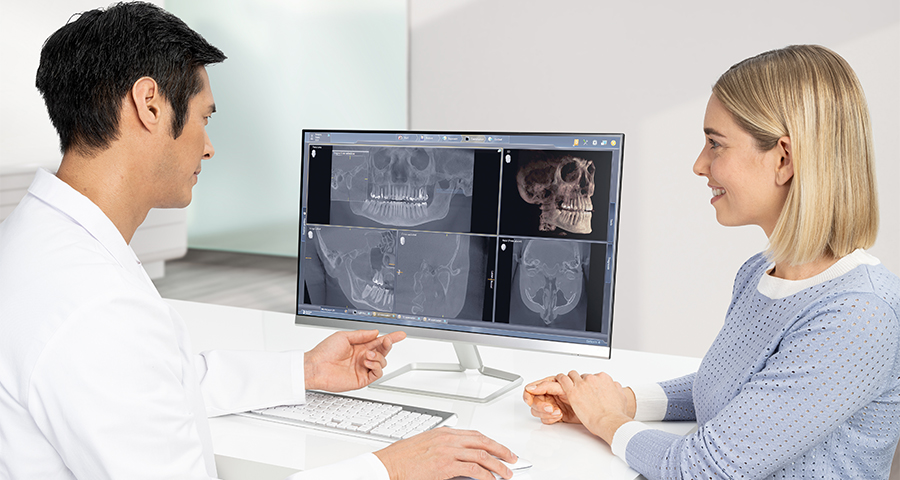
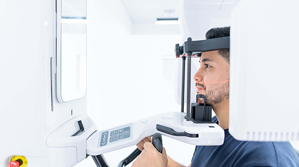
X-Ray & Digital Imaging Supplies
X-Ray & Digital Imaging Supplies
X-Ray Developing & Processing Supplies
Chairside Darkroom & Film Processor Replacement Parts
Darkroom Safelights & Filters
Developing Tank & Solution Accessories
Processor & Tank Cleaning Solutions
PSP Plate Guides & Transfer Boxes
X-Ray Cleaning Sheets & Wipes
X-Ray Developers & Fixers
X-Ray Film Carriers
X-Ray Film & Sensor Holders & Positioners
Intraoral Photography Mirrors
Panoramic X-Ray Bite Pieces & Chin Supports
Positioning Rings & Arms
X-Ray Cassettes
X-Ray Film & Digital Sensor Holder Kits
X-Ray Film & Digital Sensor Holders
X-Ray Screens
X-Ray Film, PSP Plates & Sensor Accessories
PSP Plates
X-Ray Duplicating Film
X-Ray Extraoral Film
X-Ray Film & Digital Sensor Accessories
X-Ray Intraoral Film